Figure 113 Drag The Appropriate Labels To Their Respective Targets. External acoustic meatus External auditory canal also called external auditory meatus or external acoustic meatus passageway that leads from the outside of the head to the tympanic membrane or eardrum membrane of each ear.
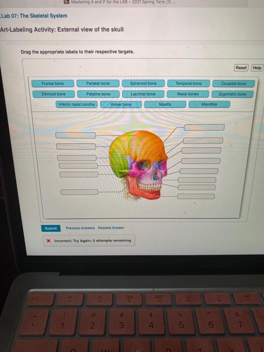
Solved Mastering A And P For The Lab 2021 Spring Term 0 Chegg Com
Bones of the Right Wrist and Hand anterior view Art-labeling Activity.
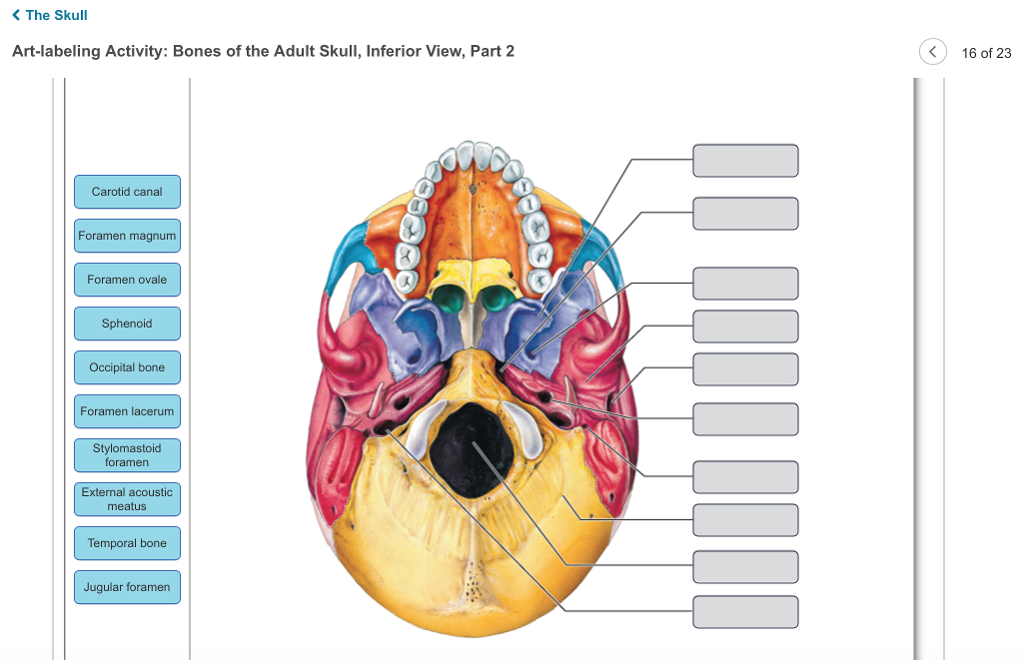
. Inferior View of the Skull Zygomatic bone Frontal bone Palatine bone Maxilla Vomer I I Foramen ovale Foramen lacerum Sphenoid bone enge ORADOS Carotid canal Zygomatic arch Jugular foramen Styloid process Stylomastoid foramen Mandibular fossa Mastold process Foramen magnum Temporal bone Occipital condyles Lambdold suture Occipital bone Inferior. View art labeling activity - the vertebral columnjpg from ANT MISC at Miami Dade College Miami. Features of the floor of the cranial cavity superior view Identify the location of the occipital bone.
Reset Help Frontal bone Parietal bone Sphenold bone Temporal bone Occipital bone Ethmoid bone Palatine bone Lacrimal bone Nasal bones Zygomatio bone Interior nasal concha Vomer bono Maxilla Mandible 000000 DID. As you read through this material identify each bone on an in. Also use the brain model as.
B The hinge joint of the elbow works like a door hinge. Posterior surface and base of the cranium Name the opening in the occipital bone through which the spinal cord passes. Posted 7 days ago Q.
YOU MIGHT ALSO LIKE. Biology Chemistry Earth Science Physics Space Science View all. The sphenoid bone is one of the seven bones that articulate to form the orbit.
Mastering A P Chapter 6 Bones And Skeletal Tissues Flashcards Quizlet Part A Drag the labels to identify the structures of a long bone. View art labeling activity - the vertebral columnjpg from ANT MISC at Miami Dade College Miami. Start studying Art-labeling Activity.
Chambers and vessels of the heart superior view of the thoracic cavity Hip bone. How many bones of the skull are considered facial bones. Key Structures of a Synovial.
In this article we shall look at the anatomy of the elbow joint. Its shape somewhat resembles that of a butterfly or bat with its wings extended. 1159pm on Friday October 6 2017 To understand how points are awarded.
Help labeling the bones and synovial joints of the body by looking at the image and providing the bone AND synovial joint type for each number. Rese Ureters External urethral sphincter Urethra Urinary bladder Trigone Rugae Levator ani muscle Internal urethral sphincter. Learn vocabulary terms and more with flashcards games and other study tools.
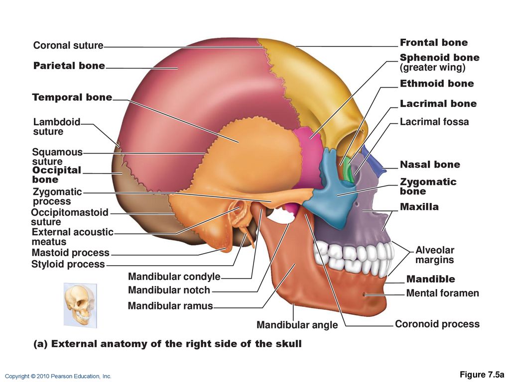
The mandible is the only movable bone of the skull besides the ossicles of the middle ear. External view of the skull Drag the appropriate labels to their respective targets. Activity 1 Identifying External Brain Structures Identify external brain structures using the figures cited.
435 Art Labeling Activity. Start studying Art-labeling Activity. English French German Latin Spanish View all.
Human skull superior view top of cranium removed. Art History Dance Film. Posterior surface and base of the cranium.
Gross anatomy of the lung right lung lateral surface Art-labeling Activity. Synovial Membrane inner- secretes produces synovial fluid for lubrication. Reset Help Brain And Spiral Cord Grative And Control Centrs Cras Nerves And Sal Nerves Commons Between The CNS And The Rest Of The Body Central Nervous System.
External view of the skull Drag the appropriate labels to their respective targets. Rotational Movements of the Joints. Frontal bone Parietal bone Sphenold bone Temporal bone Occipital bone Ethmoid bone Palatine bone.
External view of the skull. An unregistered player played the game 52. With high mitotic activity and they are the only bone cells.
Human skull inferior view mandible removed Figure 512. Rotational Movements of the Joints. It is situated in the middle of the skull towards the front in front of the temporal bone and the basilar part of the occipital bone.
BONES OF THE AXIAL AND APPENDICULAR SKELETON. The structure of a long bone humerus of arm Figure 59. Correct Spotlight Figure 82.
The cranium skull is the skeletal structure of the head that supports the face and protects the brainIt is subdivided into the facial bones and the brain case or cranial vault Figure 1The facial bones underlie the facial structures form the nasal cavity enclose the eyeballs and support the teeth of the upper and lower jaws. Figure 193 Label the different types of capillaries and their structures. Gross anatomy of the lung left lung lateral surface Art-labeling Activity.
Foramen magnum Identify the area of the occipital bone that articulates with the vertebral column. Log in Sign up. Bones of the Axial Skeleton.
Drag the appropriate labels to their respective targets. An unpaired bone of the neurocranium. After you have studied the bones in lab label the drawings as a self-test.
Anatomy of the urinary tract 18 of 24 Drag the appropriate labels to their respective targets.

Skull Lab Prep Review Flashcards Quizlet
Solved The Skull Art Labeling Activity Bones Of The Adult Chegg Com
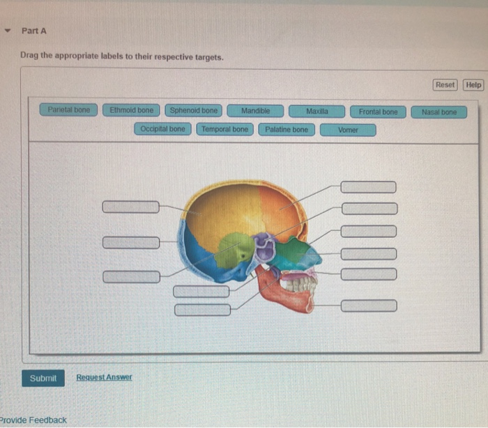
Solved Part A Drag The Appropriate Labels To Their Chegg Com
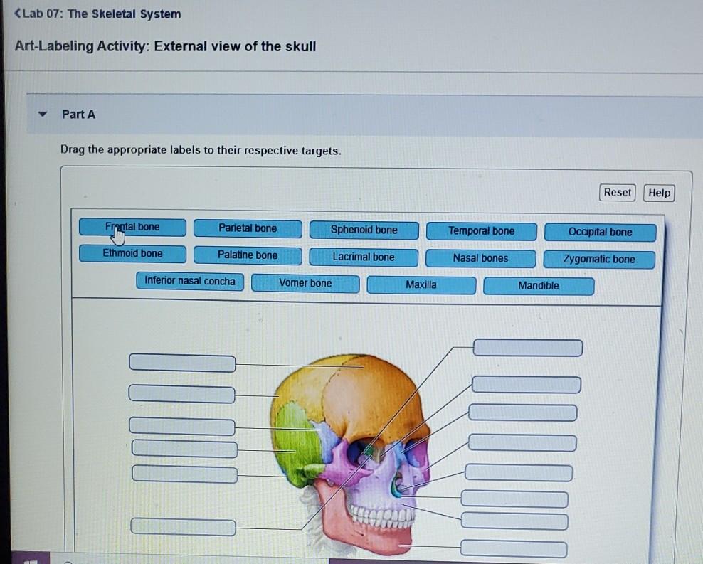
Solved Lab 07 The Skeletal System Art Labeling Activity Chegg Com

Solved Pre Lab A Labs 5 And 6 General Bone Histology And Chegg Com

Skull Lab Prep Review Flashcards Quizlet
Chapter 7 The Skeleton Shilla Chakrabarty Ph D Ppt Download

Mastering A P Chapter 7 The Skeleton Art Labeling Activity Figure 7 4a 2 Of 2 Diagram Quizlet
0 comments
Post a Comment